Immune system (part 2)
Immune system (part 2)
Dendritic cells (DC)
• are phagocytes in tissues that are in contact with the external environment; therefore, they are located mainly in the skin, nose, lungs, stomach, and intestines
• named for their resemblance to neuronal dendrites, as both have many spine-like projections, but dendritic cells are in no way connected to the nervous system
• serve as a link between the bodily tissues and the innate and adaptive immune systems, as they present antigen to T cells, one of the key cell types of the adaptive immune system


CELLS OF INNATE AND ADAPTIVE
IMMUNITY
• Lymphoid Lineage
– Large lymphocytes (large granular lymphocytes)
• Natural killer (NK) cells (CD16, CD56)
• Innate immunity to viruses and other intracellular pathogens
• Participate in antibody-dependent cell-mediated cytotoxicity (ADCC)
– Small lymphocytes
• B cells (CD19)
• T cells (CD3, CD4 or CD8)
• Adaptive immunity
– Lymphocytes refers to small lymphocytes
Lymphocytes; B cells; T cells
• B cells & T cells carry receptor molecules that recognize specific targets
• T cells recognize a “non-self” target, such as a pathogen, only after
antigens (small fragments of the pathogen) have been processed and
presented in combination with a “self” receptor called a major
histocompatibility complex (MHC) molecule
• There are two major subtypes of T cells: the killer T cell and the helper T
cell
• Killer T cells only recognize antigens coupled to Class I MHC molecules,
while helper T cells only recognize antigens coupled to Class II MHC
molecules
• A third, minor subtype are the γδ T cells that recognize intact antigens that
are not bound to MHC receptors
• In contrast, the B cell antigen-specific receptor is an antibody molecule on
the B cell surface, and recognizes whole pathogens without any need for
antigen processing. Each lineage of B cell expresses a different antibody,
so the complete set of B cell antigen receptors represent all the antibodies
that the body can manufacture
Lymphocytes
• Many types; important in
both humoral and cell-mediated immunity
• B-cells produce antibodies
• T- cells
– Cytotoxic T cells
– Helper T cells
• Memory cells


• Plasma Cell (in tissue)
– Fully differentiated B cells,
secretes Ab
• Natural Killer cells
– Kills cells infected with
certain viruses
– Both innate and adaptive
– Antigen presentation


Typical recognition markers for Lymphocytes


Comparison of T and B cells
T-cells
• responsible for cell mediated
immunity
• Life span is long
• Differentiate inside Thymus Gland
• Absence of surface antibodies
• Transformed in small
lymphocytes by antigens
• secrete Lymphokines
• sub population are Cytotoxic T,
Helper cells and suppressor cells.
• stimulate phagocytes and B-cells
into activity.
• responsible for Humoral
immunity
• Life span is short
• Differentiate inside Bone Marrow
• Surface Antibodies present
• Transformed to plasma cells by
antigens
• secrete antibodies
• sub population are memory cells
and plasma cells
• B-cells or B-lymphocytes produce
antibodies .

THE CLUSTER OF DIFFERENTIATION (CD)
• CD nomenclature established in 1982
– 1
st International Workshop and Conference on Human Leukocyte
Differentiation Antigens (HLDA) held in Paris
• protocol for identification and investigation of cell
surface molecules
• intended for classification of many monoclonal
antibodies generated by different laboratories
around the world against epitopes on the surface
molecules of leukocytes
• CD number assigned on basis of 1 cell surface
molecule recognized by 2 specific m Ab
cluster of differentiation
• The (cluster of designation) (often abbreviated as CD) is a protocol used for the identification and investigation of cell surface molecules
present on White blood cells
• CD molecules can act in numerous ways, often acting as receptors or
ligands (the molecule that activates a receptor) important to the cell
• A signal cascade is usually initiated, altering the behavior of the cell
• Some CD proteins do not play a role in cell signaling, but have other
functions, such as cell adhesion
• CD for humans is numbered up to 350 most recently (as of 2009).
• If the molecule has not been well-characterized, or has only one mAb, it is usually given the provisional indicator "w" (as in "CDw186")

THE CLUSTER OF DIFFERENTIATION (CD)
• CD markers on leukocytes
Granulocyte CD45+, CD15+
Monocyte CD45+, CD14+
T lymphocyte CD45+, CD3+
T helper lymphocyte CD45+, CD3+, CD4+
T cytotoxic lymphocyte CD45+, CD3+, CD8+
B lymphocyte CD45+, CD19+
Natural killer cell CD45+, CD16+,
CD56+, CD3-

Components of blood

COMPLETE BLOOD COUNT WITH
DIFFERENTIAL (CBC WITH DIFF)
References Ranges
Erythrocytes (RBC) 4.0 to 5.4 M/uL
Thrombocytes (Platelets) 145 to 400 K/uL
Leukocytes (WBC) 4.8 to 10.8 K/uL
Neutrophils 40 to 74 %
Band neutrophils 0 to 9
Eosinophils 0 to 6
Basophils 0 to 1
Lymphocytes 15 to 47
Monocytes 0 to 12

Other Blood Cells
• Megakaryocyte
– Platelet formation
– Wound repair
• Erythrocyte
– Oxygen transport


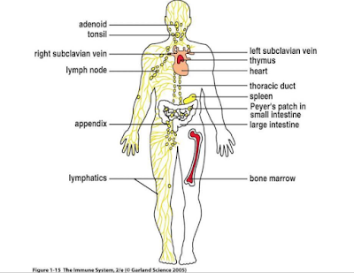
LYMPHOCYTES, LYMPHOID TISSUES
AND ORGANS
• Lymphocytes originate in bone marrow
• Lymphoid tissues and organs
– Primary
• Development and maturation of lymphocytes
• Bone Marrow (B cells) and thymus gland (T cells)
– Secondary
• Mature lymphocytes meet pathogens
• Spleen, adenoids, tonsils, appendix, lymph nodes, Peyer’s patches, mucosa-associated lymphoid tissue (MALT)
THE LYMPHATIC SYSTEM
• Lymph
– Fluid and cells in lymphatic vessels
• Lymphatic vessels
– Collect and return interstitial fluid to blood
– Transport immune cells throughout body
– Transport lipid from intestine to blood
• Lymph nodes
– Kidney shaped organs at intervals along lymphatic vessels
• Other secondary lymphatic tissues and organs

LYMPHOCYTES AND THE LYMPH
NODES
• Naïve lymphocytes circulate between blood,
lymph and secondary lymph nodes
• Pathogens from infected tissue sites are
picked up by lymphatic vessels and arrive at
closest lymph node
• T and B cells congregate at specific regions of
nodes
• Architecture and size of nodes change in
response to activation of lymphocytes


LYMPHOCYTES AND THE SPLEEN
• Spleen
– Lymphoid organ in upper left abdomen
– Functions
• Remove damaged or old erythrocytes
• Activation of lymphocytes from blood borne pathogens
• Architecture of Spleen
– Red pulp
• Erythrocytes removed
– White pulp
• Lymphocytes stimulated

SECONDARY LYMPHOID TISSUES
ASSOCIATED WITH MUCOUS
MEMBRANES
• Primary portals of entry for pathogens
– Respiratory tract
– Gastrointestinal tract
• Secondary lymphoid tissues
– Bronchial-associated lymphoid tissue (BALT)
– Gut-associated lymphoid tissues (GALT)
• Tonsils, adenoids, appendix, Peyer’s patches
• Pathogens are directly transferred across
mucosa by “M” cells


Disclaimer:
The content provided in this article is based on personal knowledge and educational background in the fields of health and nutrition. It is intended for informational purposes only and should not be considered a substitute for professional medical advice. For any health concerns, please consult a qualified healthcare provider .
The content provided in this article is based on personal knowledge and educational background in the fields of health and nutrition. It is intended for informational purposes only and should not be considered a substitute for professional medical advice. For any health concerns, please consult a qualified healthcare provider .


Comments
Post a Comment